
Quick answer. A leg ulcer is a break in the skin below the knee – most often near the inner ankle – that fails to heal, and the commonest cause is chronic venous insufficiency (poor drainage of blood up the leg veins), giving a venous leg ulcer. Because other causes – poor arterial supply, diabetes-related damage, or a mix – need very different treatment, the cause must be properly assessed – including a check of the arterial blood supply – before treatment such as compression is started.
Concerned about circulation, varicose veins or leg pain?
Message our care team on WhatsApp. Tell us what’s going on and we’ll help you find the right vascular surgeon — free, with no obligation.
Chat with our care team on WhatsAppNot sure who to see? Find the right specialist →
In a medical emergency, or for severe or sudden symptoms such as breathing difficulty, chest pain, fainting or stroke signs, call 995 immediately.
The safety point that matters most: compression is the mainstay for venous ulcers, but the artery supply must be checked first – strong compression on a leg with poor arterial flow can cause harm.
What a leg ulcer is
A leg ulcer is a break in the skin of the lower leg or foot that is slow to heal – in practice, a wound that has stayed open over an extended time rather than closing as a fresh graze would. It is common, particularly in older adults, and it matters because a leg ulcer is a sign, not a diagnosis in itself: something is stopping the skin from healing. Finding and treating that underlying cause is the key both to healing the wound and to stopping it coming back. Left unaddressed, a small break can widen, become painful, or get infected, so a leg wound that has not healed deserves proper assessment rather than repeated home dressings alone.
What a venous leg ulcer is
The leg veins carry blood back up to the heart, helped by one-way valves and the calf-muscle pump. When valves fail or veins are blocked (chronic venous insufficiency, often after varicose veins or a past DVT), blood pools and pressure builds in the lower leg. Over time this damages the skin near the ankle so a minor knock turns into an open wound that won’t heal – typically around the inner ankle, surrounded by skin that already shows venous changes. This is the most common pattern of leg ulcer, but it is not the only one.
Main types and causes in plain terms
Treatment depends entirely on the cause, so assessment comes before any bandaging:
- Venous ulcers – the most common type, caused by poor vein circulation (chronic venous insufficiency). They usually sit around the ankle and often come with swelling and skin changes; treated mainly with compression;
- Arterial ulcers – from poor arterial supply due to peripheral artery disease; often on the foot, toes or shin, frequently painful (especially with the leg raised), with cold, pale skin. These need urgent vascular assessment, and compression can be dangerous here;
- Diabetic / neuropathic foot ulcers – from nerve damage and pressure in diabetes, so the person may not feel the wound; highly relevant in Singapore;
- Mixed or other causes – a combination of vein and artery problems, or occasionally infection, poor lymph drainage, or, rarely, a skin cancer that can mimic an ulcer. These need specialist judgement.
Why identifying the cause matters
Identifying the cause is not a formality – it changes the treatment completely. Compression is central for venous ulcers because it helps the veins drain, but the very same compression can be harmful on a leg whose arteries are already struggling to deliver blood. A diabetic foot ulcer needs pressure taken off the wound and good blood-sugar control, while an arterial ulcer may need blood flow restored before it can heal at all. For that reason, an assessment of the circulation comes first, and the same wound can be managed very differently depending on what is found.
Symptoms
The core sign is an open sore or wound on the lower leg or foot that does not heal. Around it you may notice pain that varies by cause – venous ulcers often ache or feel heavy and ease with the leg raised, while arterial pain is often more severe and can be worse when the leg is elevated or at night. Other features include swelling, discharge or weeping from the wound, an unpleasant odour if it becomes infected, and changes in the surrounding skin. In venous disease that skin often shows swelling, brown discolouration, venous eczema, and hardened skin (lipodermatosclerosis) that can give the lower leg an inverted-champagne-bottle shape. In diabetes, reduced sensation can mean a wound is painless and easily missed.
Who is at higher risk
Some people are more prone to leg ulcers. Risk is higher with older age, varicose veins or a previous DVT, peripheral artery disease, and diabetes. Being overweight, having reduced mobility or spending long periods sitting or standing, and having had a leg ulcer before all add to the risk. Several of these often occur together, which is one reason a full assessment of both veins and arteries is so useful.
How it is assessed
Assessment starts with examining the wound and the whole leg, and checking both the veins and the arteries. Before compression is applied, clinicians check the arterial supply, most commonly with the ankle-brachial pressure index (ABPI) – a Doppler measurement comparing ankle and arm blood pressures. This matters because strong compression on a leg with poor arterial supply can restrict blood flow and cause harm. Interpreting the result is a clinician’s job – it is not a self-test – but the principle is simple: check the supply first, then choose the safe level of compression. Swabs may be taken if infection is suspected, and the team will look for underlying conditions such as diabetes or vein disease. An ulcer that will not heal despite good care is reviewed again for other causes, because rarely a skin cancer can look like an ulcer.
Treatment
The heart of treatment is treating the cause. For venous ulcers that means compression and elevation once arterial disease has been excluded; for arterial ulcers it means restoring blood flow; and for diabetic foot ulcers it means relieving pressure on the wound and controlling diabetes. Alongside this, good wound care matters for every type:
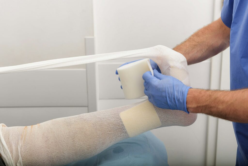
- Compression therapy (multilayer bandaging or medical stockings) for venous ulcers – the mainstay, fitted and applied by trained staff;
- Wound care and dressings to keep the wound clean and at the right moisture level;
- Leg elevation and calf exercise / walking to help venous return;
- Treating infection, providing pain relief, and managing the surrounding skin;
- Good nutrition, which supports healing;
- Treating the underlying problem – assessment of varicose veins or reflux for venous disease, or restoring circulation for arterial disease, with procedures where appropriate.
Care often involves specialist wound-care nurses and, depending on the cause, a vascular specialist, podiatry or a diabetic foot service. Healing can take time – often weeks to months – and needs consistent follow-up rather than a single visit.
Preventing recurrence
Ulcers commonly recur if the underlying problem is not managed, so prevention continues after the wound has closed. For venous disease that means ongoing compression (usually medical stockings) and careful skin care to keep the skin supple and protected. For diabetes it means daily foot care, well-fitting footwear and regular foot checks. Across all types, managing risk factors – blood sugar, weight, blood pressure, and stopping smoking – and staying active with regular walking and leg movement all help keep the skin healthy and the circulation working.
Self-care
Between clinic visits you can support healing by keeping to the compression and dressing plan your team has set, elevating the leg when resting, and walking and moving the ankle to work the calf pump. Keep the skin around the wound clean and moisturised where advised, protect the leg from knocks, and eat well to support healing. If you have diabetes, check your feet daily – using a mirror or asking someone to help for areas you cannot see – and never ignore a new blister, crack or sore. Do not apply strong compression or bandaging yourself without a clinician confirming your arteries are safe for it.
When to seek help and red flags
See a doctor or wound-care service for any leg or foot wound that is not healing, and go early if you have diabetes and notice any foot wound at all, however small. Seek urgent care for signs of infection – spreading redness, warmth, swelling, pus, a bad smell, increasing pain, or feeling feverish or unwell – and for a rapidly worsening or very painful ulcer, or any black or dying tissue. A suddenly cold, pale and painful foot may be an arterial emergency and needs immediate attention. Go to A&E or call 995 for signs of serious infection or sepsis, such as high fever, confusion or feeling very unwell. In Singapore you can start with a GP, who can refer you to a wound-care nurse or service, a vascular specialist, podiatry or a diabetic foot service, and HealthHub carries further guidance on wound and diabetic foot care.
Related vascular & circulation topics
- Peripheral artery disease (PAD)
- Critical limb ischaemia
- Carotid artery stenosis
- Superficial thrombophlebitis
- Chronic venous insufficiency
- Spider veins
- Venous eczema
- Lymphoedema
- Sclerotherapy
- Varicose veins
- Deep vein thrombosis (DVT)
- Vascular surgeons in Singapore
Sources
- NHS — Venous leg ulcers: venous cause, compression as mainstay, ABPI assessment before compression.
- DermNet — venous leg ulcer: causes, appearance, surrounding skin changes, management.
- StatPearls — venous leg ulcer: commonest leg ulcer, venous-hypertension mechanism, compression, recurrence risk.
- Wounds UK / NHS ABPI guidance — checking arterial supply before compression. HealthHub / SG diabetic-foot literature — SG context (figures vary, cited as background).