
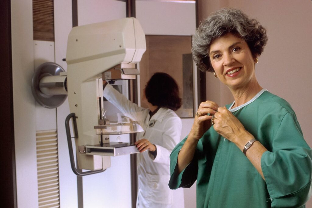
Breast calcifications are tiny deposits of calcium in breast tissue that show up on a mammogram as small bright white specks. They are extremely common, especially with age, and are usually benign. They are not related to how much calcium you eat or take as supplements. The key is the type and pattern: larger macrocalcifications are almost always harmless, while tiny microcalcifications are usually benign too but, in certain clustered or line-like patterns, can be an early marker of change in a duct that needs further assessment.
Noticed a breast change, or due for a check?
Message our care team on WhatsApp. Tell us what’s going on and we’ll help you find the right breast surgeon — free, with no obligation.
Chat with our care team on WhatsAppNot sure who to see? Find the right specialist →
In a medical emergency, or for severe or sudden symptoms such as breathing difficulty, chest pain, fainting or stroke signs, call 995 immediately.
Calcifications are read on imaging, so the “red flag” is a radiologist’s decision — attend any callback for extra views or a biopsy. Separately, always report any felt lump, skin change, or bloody nipple discharge, whatever a mammogram showed.
What they are
Calcifications are calcium deposits within breast tissue that cause no symptoms and cannot be felt. Macrocalcifications are larger, coarse white dots that are almost always benign and usually need no follow-up, while microcalcifications are tiny flecks like fine grains of salt — most are benign, but the radiologist looks at their shape and arrangement, because certain patterns can reflect activity or change within a milk duct.
Causes and who gets them
They are very common and largely age-related, becoming more frequent as women get older, especially macrocalcifications. Benign causes include normal ageing of breast tissue and blood vessels, previous injury or surgery, past infection or inflammation, fibroadenomas, cysts, fat necrosis, and calcium within milk ducts. Microcalcifications can sometimes reflect change within a duct, and in a minority a suspicious cluster can be an early sign of ductal carcinoma in situ (a very early, pre-invasive change) or, less often, early cancer — which is why the pattern matters.
How they are found
Calcifications cause no symptoms — you cannot feel them and they cause no pain or lump — and they are almost always found incidentally on a mammogram, including a routine screening mammogram. This is one reason mammographic screening can pick up very early changes before anything is felt; the screening programme itself is covered in our breast-screening guide.
Red flags — a radiology decision
Because calcifications are read on imaging, the decision about whether a pattern is benign or needs further work-up is the radiologist’s. Generally reassuring patterns are scattered, widely spread, coarse calcifications, round or uniform specks, and calcification in blood-vessel walls. Patterns that prompt further imaging or a biopsy are microcalcifications that are tightly clustered, in a line, or branching or segmental (suggesting they lie along a duct), or that are new, increasing or irregular in shape. The finding is graded using BI-RADS categories in the report — broadly, normal or benign categories need no action, a “probably benign” category usually means a short-interval follow-up, and a suspicious category usually means a biopsy. Separately, always report any lump, skin change, nipple change or bloody nipple discharge to a doctor regardless of what a mammogram showed.
How they are assessed
Assessment uses additional magnification or spot-compression mammogram views to see the calcifications more clearly and judge their shape and grouping, and comparison with previous mammograms — stable calcifications over years are reassuring, while new or increasing ones get more attention. Ultrasound is limited for the calcifications themselves but may assess surrounding tissue. If a pattern is suspicious, a biopsy is done, typically a stereotactic (mammogram-guided) or vacuum-assisted core biopsy that samples the exact spot, sometimes leaving a tiny marker clip behind.
What happens next
For benign macrocalcifications, no treatment is needed and often no special follow-up beyond routine screening. “Probably benign” calcifications are usually monitored with a repeat mammogram, commonly after about six months, to confirm they are stable, rather than biopsied straight away. Suspicious calcifications are biopsied to get a diagnosis, and if the biopsy is benign you return to routine follow-up. The key reassurance is that most calcifications, including many microcalcifications, turn out to be benign — being called back for extra views or a biopsy does not mean you have cancer, it means the radiologist wants to be certain and, if there is any early change, to catch it while it is most treatable. It therefore matters to attend any recall rather than skip it, and to keep any previous mammogram films or reports available, since comparison over time is one of the most powerful tools for deciding whether calcifications are harmless. There is nothing you can do through diet, supplements or lifestyle to dissolve or prevent calcifications; the useful action is simply to keep attending routine screening at the recommended intervals so that any pattern which does matter is picked up and assessed early, when it is easiest to treat. If you move clinics or change your regular mammogram provider, ask for your prior images to be transferred so the new radiologist can compare rather than start from scratch.
When to see a doctor
Calcifications are usually managed through the mammogram-reporting and recall process, so attend any recommended callback, extra imaging or biopsy appointment. Independently, see a doctor promptly for any felt lump, skin or nipple change, or bloody or spontaneous nipple discharge — these are not 995 emergencies but should be assessed without delay.
Related breast-health topics
- Fibrocystic breast changes
- Intraductal papilloma
- Breast abscess
- Galactorrhoea (milky discharge)
- Mammary duct ectasia
- Phyllodes tumour
- Breast self-examination
- Inverted nipple
- Fat necrosis of the breast
- Breast cancer screening
- Breast surgeons in Singapore
Sources
- Breastcancer.org — Calcifications on a mammogram: breastcancer.org
- American Cancer Society — Understanding Your Mammogram Report (BI-RADS): cancer.org
- SingHealth — Breast Cancer (imaging and biopsy pathway): singhealth.com.sg
- Breast Cancer Foundation Singapore: bcf.org.sg